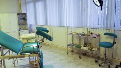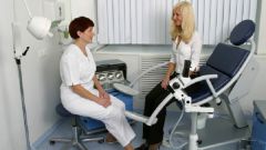
Instruction
1
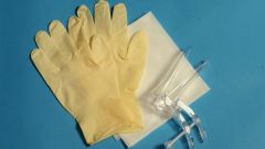
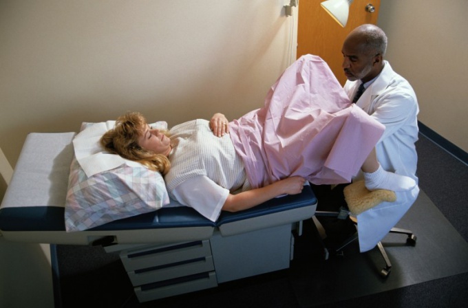
Wash your hands with soap and water, wipe with a towel. Can use special medical solutions that kill bacteria. Wear disposable gloves. Inspection start with the pubis. Note its form, the nature of volosistoe (male type, female and mixed), the presence and magnitude of the subcutaneous fat layer. Inspect the inner surface of the thighs on the subject of eczema, warts.
2
Note the large and small labia, their size, presence of tumors, ulcers. In the anus may be hemorrhoids, prolapse of the mucosa, fissures, ulcers.
3
Spread the fingers of the left hand labia, look closely at their color, ulceration, the mucous membranes, pigmentation. Inspect the clitoris, its shape, size, whether there are any anomalies in development. Examine the orifice of the urethra, polyps, condition of mucosa, note the nature of the discharge. At the same time see if there are signs of infantilism.
4
Guide the collection of material for bacteriological, cytological, histological and microscopy. An internal study carried out with the help of mirrors, as preliminary inspection of the thumb can injure the vagina and the mucous membrane of the cervix, change the character of vaginal secretions, making unreliable the results of the inspection.
5
Take vaginal speculum, determine the status of the vaginal walls, color of mucosa, presence of the growth, tumors, etc., Examine the vault and the cervix in nulliparous round the external opening of the cervical canal, giving birth the same - it is in the form of a transverse slit. Note the pathological changes, presence of erosions, fractures, dysplasia, endometriosis submucosa, and others.
6
Conduct a combined examination. Parted fingers labia, pay attention to the width of the entrance to the vagina, the elasticity of its walls. The other arm of the lock through the abdominal wall of the investigated organ (uterus, appendages), try to probe a particular area of the pelvis.
7
Do not press on the area of the urethra, the clitoris, the anterior vaginal wall. Fingers must slide along the rear wall of the vagina. Enter them deeply, to study the structure of the mucosa, the presence of bartholinitis, partitions. Find the magnitude of the cervix, the shape, the size of the external uterine weight, its disclosure in ICN, scarring after childbirth, tumors.
8
Proceed to vaginal-brunetoochka the study, which will help to determine the position, size and shape of the uterus, condition pelvic peritoneum. The normal uterus is pear-shaped, as if tapered from front to back, its surface is smooth. By touching it shifts smoothly. Physiological reduction of the uterus and occurs in menopause. Pathology is, in particular, infantilism, and atrophy of the uterus.
9
Start your research of the appendages (ovaries and fallopian tubes) with the inspection. Unmodified pipe is soft and thin, they don't usually be felt, as well as the appendages of the uterus, ligaments. Sometimes the pipes are soldered spikes. Ovaries well be felt, they are quite agile and sensitive. When the study through the vagina is impossible, for example, the virgins, examine the patient rectally.
10
Conduct a rectal examination on the gynecological chair in gloves or fingertip, lubricated with a special tool. You can use the enema. Such a study is shown in the suspicion of the presence of changes in the structure or pathology in the vaginal wall.