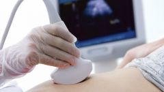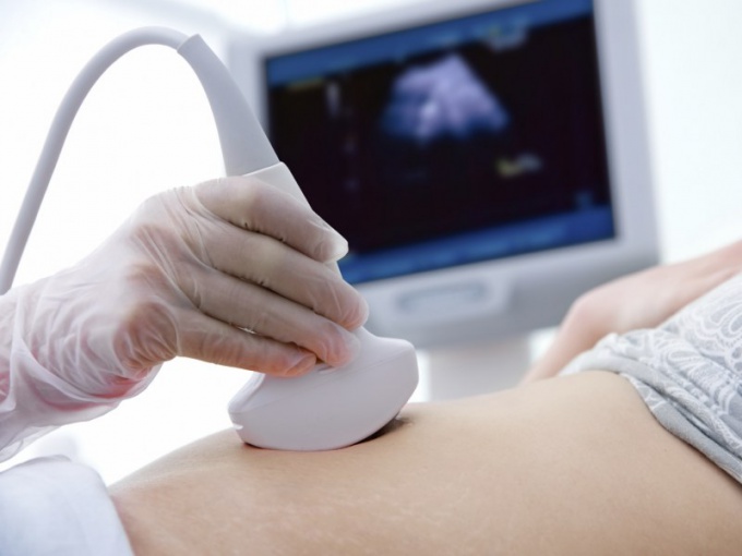
Instruction
1
The primary diagnostic goal of the first ultrasound to confirm intrauterine pregnancy, to determine the number of live fetuses in the uterus and to exclude non-developing pregnancy. The doctor can see the embryo, localization of the implant, which indirectly allows to judge about the dangers of low attachment of the placenta and the likelihood of its detachment. In this period, the expert can still conduct a survey of other pelvic organs. Always checks the condition of the ovaries to rule out cysts and other tumors. Features of the structure of the uterus is also recorded.
2
Specialist pays much attention to the yellow body (the Protocol can be specified - the yolk SAC), it is temporarily existing body which ensures development and nutrition of the embryo up to 12 weeks. After this period, it is not required and the corpus luteum decreases in size. The peculiarities of the corpus luteum can be determined missed abortion. At this period a clearly visible chorion is the embryonic sheath, which develops into the placenta.
3
CTE (coccyx-parietal size) measure necessary. These data allow to judge about both the pregnancy and on fetal health. NT (nuchal translucency thickness) is one of the most important indicators. When properly conducted study NT very reliably for the diagnosis of chromosomal disorders, particularly down syndrome.
4
Measure the size of the nasal bone, this size can be indirectly judged from the presence of down syndrome in the fetus. Syndrome "butterfly" indicates that the bones of the cranial vault are positioned correctly and visually look in the form of butterfly wings. Be sure to define anthropometric indices and heart rate (HR), along with heart rate, the doctor looks at the characteristics of blood flow.
5
All survey data should be interpreted collectively, not individually. Since this period, the growth of the fetus can be intermittent and often the doctors with inadequate experience may incorrectly diagnose developmental delay. Since for each pregnancy individually - the individual and the size of the fetus. Doctors consider it, and compare with the average and marginal population indicators.
6
Serious malformations, malformations incompatible with life at this period is almost always overlooked. Less complex malformations, the doctor may only suspect, but not always. Sex determination in this period is possible but depends on many factors, and therefore it is determined 20 weeks on the second ultrasound.
7
You must bring your passport, policy OMS and the direction of a physician, in some consultations, an ultrasound is performed right in the doctor's office, others have a private diagnostic centres. Must bring sheets or a diaper, which you will lie, and the napkin to wipe off the stomach from the gel. Because the first ultrasound is sometimes carried out through the vagina - it is better to wear a skirt, not to undress completely.
8
Today, more than 80% of pregnant women covered by stringbean study. Ultrasound is safe, his credibility is very high. A woman can abandon diagnosis, but doctors recommend it in all cases. Even if you are ready to bear a child with the condition, there will be enough time to prepare for his upbringing.