Purpose ultrasound of the fetal heart
Due to the method of ultrasound examination to identify and diagnose in the early stages of cardiovascular disease child. It is necessary to ensure that, in the case that the child's body after birth will not be able to function independently, to provide him necessary assistance and, if necessary, to prepare medicines and the incubation box.
The structure of the heart in a child who is in the womb of the mother and newborn child are significantly different. It is important that the specialist have identified abnormalities in the structure of the heart at an early stage and, if necessary, advised in time to terminate the pregnancy or do heart surgery on unborn child.
Procedure ultrasound
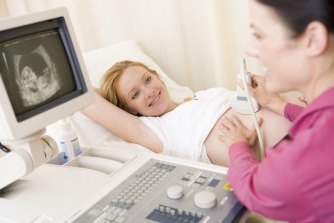
Procedure ultrasound like all future lobes, because it requires no special training and does not deliver discomfort. During the ultrasound the doctor mounts on the belly of a pregnant woman the transducer, which produces ultrasonic vibrations deep into the body. These oscillations are faced with internal organs of the mother and the child and by the child's body. Then wave back to the sensor, which sends the received signals to the analyzing device. The doctor processes the information received and only then make any conclusions.
Modern devices are capable of scanning the entire structure of the heart during fetal development. The definition of this structure is the main component of a standard procedure, ultrasound during pregnancy.
There is also transvaginally method for ultrasound. In this way, the sensor is inserted in the vagina and not cause any pain or discomfort.
Heart ultrasound in the first trimester
During the first trimester due to the ultrasound the baby's heart, the determined heart rate. This indicator makes clear to the doctors how well the child feels in the womb. The norm is 140-160 per minute. A deviation from the norm is a good reason for the risk of development of hypoxia and other violations.
Heart ultrasound in the second trimester
At 14 weeks, when comes the second trimester of pregnancy, the baby's heart is similar in structure to the heart of an adult. Beginning to take shape similarities. To give the best estimate of the development of the child's body, heart exam is required at 26-28 weeks of gestation, when they become visible elements of the structure of the heart valves and walls, and much more.
Heart ultrasound in the third trimester
As strange as it may seem, but a survey of organs of the child becomes less effective. The results in the survey this period may be false, it is therefore necessary to undergo an ultrasound procedure on time.