What you need to do an ultrasound of the kidneys and bladder?
The procedure ultrasound of the kidneys and bladder is also called echography or analysis, which involves the use of sound waves to create images of organs. During the ultrasound a fixed image is displayed on the screen of the device. Examination of the kidneys and bladder performed for an accurate diagnosis, tracking treatment and monitoring after surgery.
Thanks to the results of the ultrasound doctors have the opportunity for early diagnosis of urolithiasis, detection of tumors of any nature, anomalies of the bladder, kidney dvoenie.
The procedure of ultrasound provides accurate information about the functioning of the organs. Diagnost-uzist may reveal the following:
• congenital abnormalities;
• increased renal size;
• organ damage;
• the presence of cysts and tumors;
• complications of infectious urogenital diseases.
Do you need special preparation for ultrasound of kidneys and bladder?
Ultrasonography of the kidneys do not need special training because of their excellent imaging. To diagnosis bladder was full, before the procedure it must be tight to fill.
The safety of the procedure ultrasound of the kidneys and bladder is determined by the absence of harmful radiation on the organism and chemical damage to the skin in the course of its implementation.
The bladder is as follows:
• 1 liter of fluid (not soda) drink 2 hours before procedure ultrasound;
• refrain from voiding is allowed for 4 to 6 hours;
• taking diuretics is 1.5 hours prior to the procedure in the absence of renal disease and the cardiovascular system.
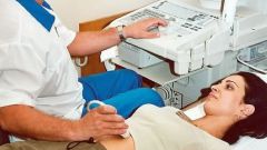
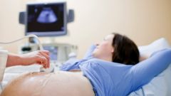
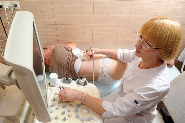
What happens in the office when ultrasound examination of the kidneys and bladder
Examination of the patient spends the technician, he is a specialist in ultrasonic echography. As beliefs in the correctness of the preparation of his patient, he offers him to change into a hospital gown. Such clothing makes it easier to perform the procedure, providing the ability to adjust the clothing based on the needs survey.
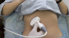
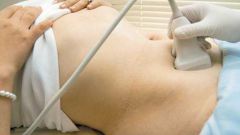
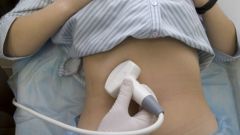
To examine the kidneys and bladder, the diagnostician would be in a dark room, since these conditions contribute to a more distinct visualization of the display bodies on the screen. The procedure begins with the application and distribution of a special gel on the patient's abdomen. Next, the technician moves the ultrasonic transducer to the body and displays the image on the screen. If it finds some images are important for the subsequent diagnostic procedures that will save them.
Ends the procedure ultrasound of the kidneys and bladder removing the gel from the skin and dressing the patient. The procedure generally takes 30 to 40 minutes. The survey results are given to patient on the hands within 1-2 days.