Instruction
1
To begin, rate the quality of the pictures. To the description, as a rule, we accept only clear photos. This means that the picture should be no blurry outlines of bodies. That is, all limbs should be removed very clearly. Only on this picture you can see the deviations from the norm.
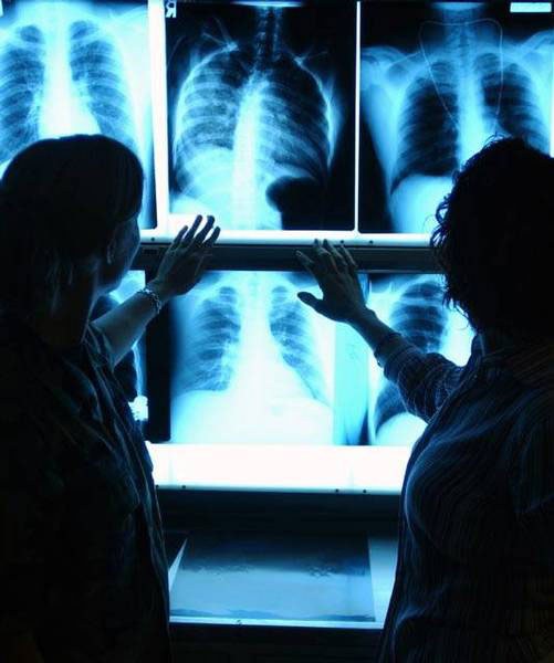
2
Be sure to choose the correct lighting. It is best to look at the pictures on the background of the fluoroscope. But if you don't have it at hand, you can use any other sufficiently bright illuminated light in a darkened room.
3
Carefully review all the details. If you have a fracture or a crack on the x-ray is necessarily affected. For example, a crack will appear as a small thin snake at the site of injury. If there is a fracture, then the picture you will notice the displacement of the bones or parts of one of them.
4
If you want to know if you can see in the picture any disease that occurs deep in the body, for example, internal haemorrhage, tuberculosis, pneumonia, etc., look for darkening. Bronchitis typically affects light. It will be seen in the picture as a darker plot.
5
Tuberculosis in the images will appear as small focal changes, i.e. darkening of size 2-3 mm. They can be both single and plural. By their number and localization can determine the degree of development of the disease. The more foci, the neglected disease.
6
X-ray allows you to see what is the situation in root canals. For example, you may be able to see the location of the material in the treated toothH. If more than a bright spot down to the end of the root, then the seal is right. If light spot is discontinuous, so should perelechivat teeth.
Note
Any the have to comment on the doctor. You can learn it only out of curiosity. The diagnosis is put only by a specialist.
Useful advice
Very carefully study the. Not to see the presence of pathology, you need to carefully measure and to see all the available dimming and other deviations from the norm.