The symptoms of low and high ICP
Increased intracranial pressure is characterized by increased amounts of fluid in the brain upon receiving a traumatic brain injury or an infection (encephalitis, meningitis). The person feels like something heavy is pressing on the head in parietal area. In the morning there is often nausea, vomiting. With a sharp forward bends noted dizziness, double vision. After a quick walk or climbing the stairs quickens the heartbeat, felt the lightheadedness. Before bed possible the heat in his temples, in the absence of high fever, General body tension.
In infants the most common cause of raised intracranial pressure is hydrocephalus - accumulation of large amounts of fluid in one of the cavities of the brain and swelling of the surrounding tissues. The size of the head disproportionate to the body, the skull becomes pear-shaped.
The decrease in intracranial pressure occurs due to trauma or wrong brain structure in violation of the integrity of bone structure in which there is leakage of brain fluid. One of the reasons is also uncontrolled long-term use of diuretics. Reduces the pressure of a pinched cervical vertebrae and venous dysfunction (vasoconstriction).
Symptoms of low intracranial pressure are: weakness, fatigue, irritability, drowsiness. People can sleep 8-10 hours and feel overwhelmed. Heaviness in the head is felt not from above but from the sides, as if head squeezed in a vise. There is often disturbance of respiration and lowering blood pressure.
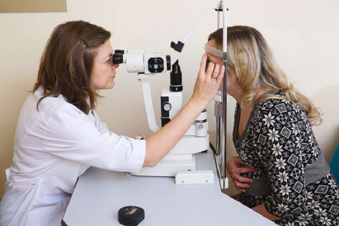
Measurement of intracranial pressure
To know the measure intracranial pressure in newborns is possible with the help of neurosonography - ultrasound of the brain. Adults due to the abundance of hair on the head, this procedure cannot be performed, so when the appropriate symptoms of the patient, the therapist prescribes a comprehensive examination.
You first need to pass the examination of the fundus by an ophthalmologist. The patient in the absence of contraindications instilled into the eyes 1-2 drops of a solution of scopolamine (0.25 percent), or homatropine (1%) for pupil dilation.
The study is conducted with the electric Ophthalmoscope or manually through the use of a special magnifier and Ophthalmoscope mirror. Electric device does not require any additional tools and allows you to get as close to the eye of the patient, and the study is carried out manually in a darkened room at a sufficient distance.
A mirror set in front of the right eye of a doctor sitting at a distance of 40-50 cm from the subject. Source of light (frosted electric lamp 60 - 100 W) is located behind and to the left of the patient, as when viewed in transmitted light. After obtaining a uniform illumination of the pupil, the researcher puts a magnifying glass (usually at 13.0 diopters.) 7-8 cm in front of the eye of the patient, resting a finger to his forehead. The pupil of the researcher, the hole mirror, the center of the magnifier and the pupil of the examinee must be on the same line.
When fundus examination optometrist draws attention to the optic disc and the vascular condition of the retina. Of increased intracranial pressure signal extended tortuous vessels and change the color, contours and fabric drive.
If there is any suspicion the patient should be referred to a neurologist. A neurologist may prescribe an MRI - magnetic resonance imaging of the brain, rheoencephalography and duplex scanning of brachiocephalic arteries, which are responsible for cerebral blood flow. For conducting any research, the patient must have a medical card, the data on the previous survey, cotton pajamas or a t-shirt.
Since the method of magnetic resonance imaging-based visualization of body cavities when the absorption of the radiation by the tissues of electromagnetic waves, in the study it is necessary to remove jewellery (earrings, chains, necklaces, piercings, etc.), watches, phone, magnetic card. Clothes should not be of metal goods (buttons, buckles, clasps).
The patient in the supine position is placed on the sliding table in a kind of capsule of cylindrical shape, surrounded by a circular magnet. On his head is worn a special helmet. Around the head of a special device - a coil receiving and emitting radio waves. During the procedure, the unit is very noisy, so for the convenience of the patient offer headphones with pleasant music. The average MRI procedure lasts about 45 minutes.
Contraindications for MRI: weight over 150 kg, the presence of metal in the body (pins, bullets, shrapnel, pacemaker, artificial blood vessels and heart, vascular clips, etc.), claustrophobia, pregnancy, a serious condition of the patient (patients on stretchers and gurneys).
Rheoencephalography - a method for registration of changes of electrical resistance of the brain and soft tissues of the skull by passing through them weak alternating current of high frequency, which does not felt. The patient is seated in a comfortable chair, placed on the head of the sucker, which joins the transaction. The light in the room turns off and on the observed direct light pulses of different frequencies. To the beat of a pulse wave occur periodic current oscillations, which after appropriate amplification can be displayed graphically in the form of a pulse of oscillations of a complex electric resistance of the rheogram.
Duplex scanning of brachiocephalic arteries is ultrasound common, external, internal carotid and vertebral arteries in the neck. The procedure takes about 5-10 minutes and allows you to visually assess the condition of the vessel width, rate of blood flow, clearance, etc.
The most accurate method for determining intracranial pressure is considered a surgical insertion of a needle into the spinal canal. However, in this procedure, few addressed, because the incorrect insertion of the needle can result for the patient pinched nerve endings of the vertebrae and subsequent disability.