Instruction
1
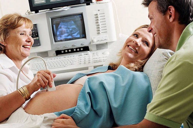
Before the deadline in 3 weeks, the doctor when using ultrasound can only see the intended site of implantation of a fertilized egg. This area has a different structure and has different degree of reflection of ultrasonic waves. Diagnostic and practical value of such research is zero, as the conclusion is not taken into account, even if the woman decides to terminate a pregnancy, choosing medical abortion. The fertilized egg can be clearly seen on the 5th day of menstruation or at the concentration of 1500 IU HCG, if you do transvaginal ultrasound sensor, and when the concentration of HCG 4500-65000 ED when abdominal examination. To 6 weeks pregnancy and her period is determined by measuring the average inner diameter of the ovum. This is the most accurate study, because the size of the ovum depends on the date of conception, and individual characteristics, hereditary factors and the structural features of the mother's body is still irrelevant.
2
Within 6 weeks, the doctor measures kopcik-parietal size, but may be error for 3-5 days, which does not affect the pregnancy tactics. Approximately 4-5 weeks you can hear the heartbeat that tells about normal developing pregnancy. However, if there is no suspicion of ectopic pregnancy, missed abortion, or on such a dangerous pathology like molar pregnancy, doctors do not recommend to do an ultrasound. The first screening ultrasound is done in the period of 11-13 weeks, it has the greatest diagnostic value. After 12 weeks, the accuracy of determination of time by means of ultrasonography is rapidly declining, and it is used only in cases where the woman doesn't know when you got pregnant or did not suspect pregnancy. Measure separately size, circumference of head, chest, abdomen and thigh length.
3
After 20 weeks with information on pregnancy the doctor is examining the placenta, cardiovascular and nervous system of the fetus. The anthropometric data may not be informative because of the pregnancy pathologies, pathologies of the fetus and the individual characteristics of the pregnant woman and the fetus. The difference in 2 weeks for the term of 20 weeks is considered a normal variant, with 30 weeks of the inaccuracy of the ultrasound method can take up to 3 weeks. The accuracy of ultrasound diagnosis may depend on the level of training of doctor, type of equipment and the individual characteristics of the patient. So, overly-developed subcutaneous fat layer may distort the result. If you need a very early diagnosis of pregnancy, it is best to take a blood test on the concentration of HCG as early pregnancy ultrasound is not always accurate.