Instruction
1
Every pregnant woman wants to hear the sound of the heart of their unborn child. Because the fetal heartbeat is considered a major sign of the vitality of the embryo. If the heart rhythm is correct, it's safe to say that the child is healthy and has no abnormalities. If the fetus ' heart completely stops beating, it generally indicates either a miscarriage or intrauterine asphyxia and other anomalies. Therefore, an important part of pregnancy is listen to the heartbeat and movements of the fetus. For monitoring cardiac activity of the child used ultrasound (ultrasound), Echocardiography (echocardiography), auscultation (listening with a stethoscope, cardiotocography.
Already the fourth week of pregnancy the embryo begins to develop the heart. At first it is a hollow tube, and then, closer to 6-8 weeks of pregnancy, it becomes similar in shape to the adult heart. During this period the heart begins to perform its functions, and the embryo receives nutrients through the primary blood vessels. Closer to the third month of pregnancy the placenta is formed, through which the exchange of nutrients between mother and child comes the so-called placental period, which lasts until the onset of childbirth.
Already the fourth week of pregnancy the embryo begins to develop the heart. At first it is a hollow tube, and then, closer to 6-8 weeks of pregnancy, it becomes similar in shape to the adult heart. During this period the heart begins to perform its functions, and the embryo receives nutrients through the primary blood vessels. Closer to the third month of pregnancy the placenta is formed, through which the exchange of nutrients between mother and child comes the so-called placental period, which lasts until the onset of childbirth.
2
In the first weeks of pregnancy monitoring cardiac activity of the embryo can be carried out only with the help of ultrasound. In the first 5 - 8 weeks the fetal heartbeat is 110-140 beats / minute, and further, since the second trimester, the heart rate accelerated to 160 beats per minute. Output heart rate outside this range indicates the presence of pathology. When heart sounds are not listened at all, it says that the pregnancy is not growing, or there is a failed miscarriage.
The pregnancy and external factors (heat, cold) have a great influence on the heart rate of the fetus. Diseases of the mother and her lifestyle also play a crucial role in the life of an embryo. Often the rhythm slows down due to a variety of heart defects of the fetus. This is especially noticeable in late pregnancy. If there is suspicion of a malformation of the heart of the embryo, the gynecologist sends a pregnant woman into a more detailed examination by echocardiography and cardiotocography.
The pregnancy and external factors (heat, cold) have a great influence on the heart rate of the fetus. Diseases of the mother and her lifestyle also play a crucial role in the life of an embryo. Often the rhythm slows down due to a variety of heart defects of the fetus. This is especially noticeable in late pregnancy. If there is suspicion of a malformation of the heart of the embryo, the gynecologist sends a pregnant woman into a more detailed examination by echocardiography and cardiotocography.
3
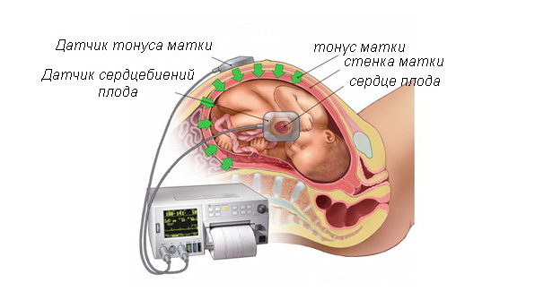
Auscultation (listening to fetal heart), unlike ultrasound, is performed later in pregnancy. For this fetal stethoscope is used, differing from ordinary fact that it has a wide bell. This stethoscope to listen not only heart sounds of the fetus, but the child's movements and sounds created by the body most pregnant women: uterine murmur, heart rate, bowel function, breathing.
Through auscultation, it is also possible to draw a conclusion about the presence in the child's development of certain pathologies and to detect multiple pregnancies, determine the position of the fetus in the uterus, depending on which is changing the place where the heart sounds are listened in the best way. If the baby is head down, heart tones are best auscultated below the umbilicus. In case, if the fetus is breech, the heartbeat can be heard at or above the navel. In addition, if the child's head is flattened, and the breast pressed to the wall of the uterus, the heart can be heard more clearly than in the flexed position of the fetus, when the child touches the back wall of the uterus.
Auscultation as ultrasound helps to monitor the pregnancy and identify any defects and diseases of the fetus. It also defines indicators of the progress of the pregnancy, possible risks to the mother. In the parameters auscultation gynecologist can conclude how beneficial is pregnancy, and, in the case of any violation, to promptly eliminate it.