You will need
- The direction of the survey.
Instruction
1
You will be given x-rays of the esophagus. In conventional x-ray the esophagus is not visible, so you will drink a solution of barium sulfate that does not transmit x-rays. In the picture you will see the relief of the esophagus.
2
For the diagnosis of diseases of the esophagus esophagogastroduodenoscopy is often used. During the procedure, immediately examined the entire esophagus, stomach and duodenum. You will have a local anaesthetic and examine all the organs using an endoscope with illumination. The image is viewed through the monitor.
3
If necessary, do a biopsy and histology. To do this, take pieces of cloth, a doctor and a pathologist will conduct a study under the microscope. This examination reveals a malignant tumor and allows timely to detect changes that precede cancer.
4
Also, you can assign endoscopic optical coherence tomography. The survey is conducted using an endoscope with an optical sensor which receives the reflection of the esophagus and transmits to the computer monitor.
5
A blood test for tumor markers is not always possible to obtain an accurate diagnosis, cancer of the esophagus is characterized by a increase of the markers CYFRA 21-1, TPA, SCC, and they are present in the blood only in 40-45% of patients when the disease has gone too far.
6
Computed tomography done if the previous methods of examination do not allow to make an accurate diagnosis. The method allows to comprehensively examine the oesophagus, to define clear boundaries between inclusions and lesions.
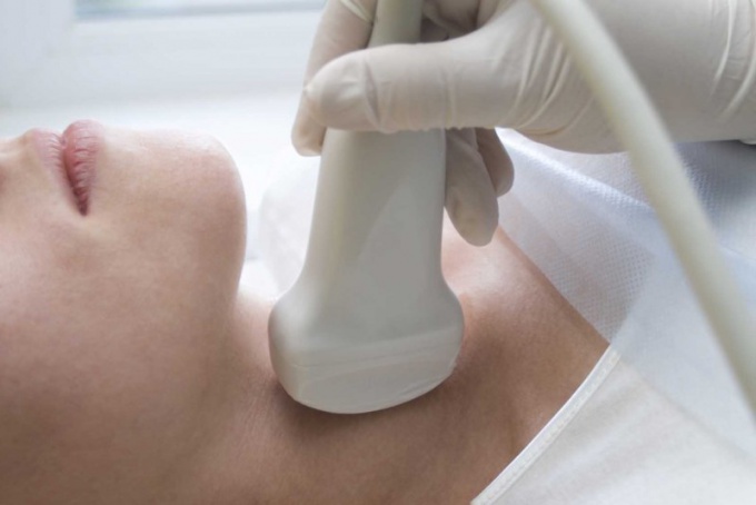
7
Ultrasound is assigned to all patients to view the structure of the tissue of the esophagus.
8
Esophagoscopy is performed for suspected cancer. Inside of the esophagus enter the sensor and inspect all the walls, if necessary with the help of this method, take the material for examinations not only of the tissues of the esophagus, but also closely located lymph nodes.
9
When performing bronchoscopy examines the entire esophagus, vocal folds, windpipe and bronchial tubes.
10
Tomography positron emission is used to detect all focal changes of the esophagus. In the vein injected radioactive glucose, which immediately accumulates in cancer cells. Using a scanner examines the degree of damage of the esophagus.
11
Final diagnosis the doctor can only put the result of a comprehensive study.