What is ultrasound?
Ultrasonography (us) is one of the modern methods of diagnostics in medicine, allowing to visualize the organs using ultrasonic waves. For visualization of the interior of the human body using acoustic waves of high frequencies are not audible to the human ear. They painlessly penetrate the body without damaging cells.
Ultrasonic waves are safe, in contrast to x-rays. In addition, does not require introduction of fluid in the body to create contrast.
What organs can be explored through an ultrasound?
Pediatric ultrasound is used for examination of the brain in neonates and infants. In gynecology using ultrasound to examine reproductive organs of women, and to monitor pregnancy.
Ultrasound is widely applied in the study of the thyroid gland, adrenal glands, kidneys, ovaries, testes, liver, gall bladder, pancreas, bladder, prostate gland and other internal organs. Every body reflects the ultrasound, making it possible to determine its structure.
How to prepare for an ultrasound?
For an hour and a half before the procedure it is advisable to drink a liter of mineral water or unsweetened tea to during the study, the bladder was filled. Before the test - do not smoke because the smoke distorts the image.
For the quality of the ultrasound examination of abdominal cavity a few days before the deadline of the procedure should be excluded from the diet meals that trigger gas formation in the intestine. In the morning an ultrasound of the abdomen done on an empty stomach, and if the procedure is scheduled for the second half of the day, you can eat easily digestible Breakfast.
Ultrasound other soft tissues (muscles, lungs, Central nervous system and organs of the neck) usually does not require special training.
Procedure ultrasound
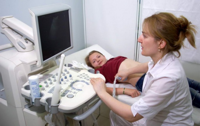
An important part of the ultrasound machine is a head in which the ultrasonic Converter. The image of the selected organ or tissue appears on the monitor due to the fact that the acoustic pulse is replaced by electric.
Before the study is required to reveal the corresponding part of the body. Then the doctor puts on the patient's skin with a special gel, which, by suppressing air bubbles, ensures a good contact of the head with the body. Moving the head in a specific area of the body, the doctor receives an image of the organ being examined on the monitor screen. The study can be repeated multiple times, even after short periods of time.